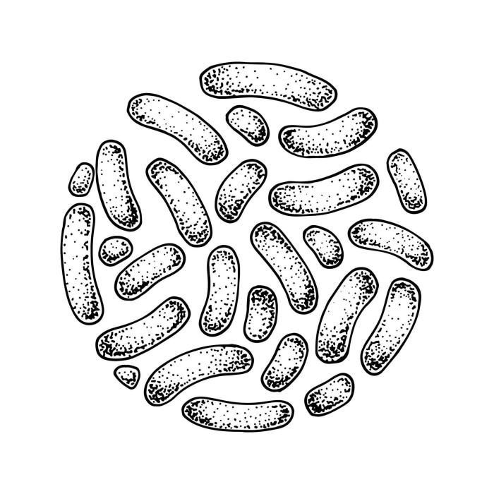
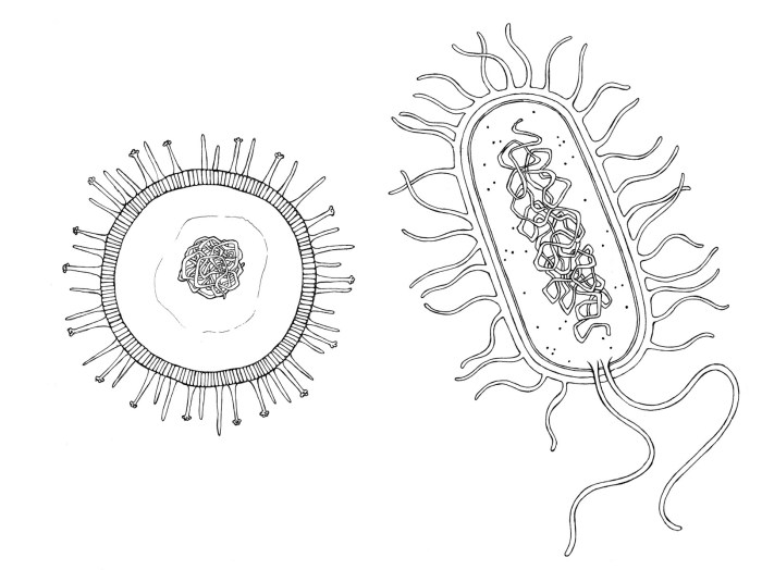
The Cold Drawing Process

Black and bacteria cold drawing easy – Embark on a creative journey into the microscopic world! Cold drawing, a technique using pencils on paper, allows for the precise and detailed rendering of subjects like bacteria. This process emphasizes skill, patience, and an eye for detail, transforming simple strokes into complex forms. Let’s explore the materials and techniques that will bring your bacterial illustrations to life.
Mastering cold drawing involves understanding the interplay between materials and technique. The right tools significantly impact the final outcome, allowing for a range of textures and shades. Careful observation and practice are key to capturing the subtle nuances of form and structure.
Material Selection
Selecting the appropriate materials is crucial for a successful cold drawing. A smooth, high-quality drawing paper provides a superior surface for detailed work. Consider using cartridge paper or Bristol board, known for their texture and ability to handle layering. For pencils, a range of grades is recommended, from a hard 2H for light sketching and fine details to a soft 6B for achieving deep blacks and shading.
A kneaded eraser is essential for lifting graphite without damaging the paper, allowing for adjustments and corrections throughout the drawing process. A sharpener, preferably a hand-held one for precise control over the pencil point, completes your essential tool kit.
Unleash your inner artist with simple black and bacteria cold drawings! The seemingly mundane can become a masterpiece with practice. For a fun time-management exercise, try incorporating elements of time into your artwork, perhaps by referencing a helpful tutorial like this one on clock easy drawing with dates. Then, return to your black and bacteria drawings, applying newfound precision and patience.
Every stroke is a step forward on your artistic journey.
Pencil Techniques for Rendering Texture and Depth
Several pencil techniques are employed to create the illusion of three-dimensionality and texture in a drawing. These techniques manipulate the density and direction of pencil strokes to achieve varied levels of shading and contrast.
Hatching involves applying a series of parallel lines in a single direction. The closer the lines, the darker the tone. Cross-hatching builds upon hatching by adding another layer of parallel lines at a right angle to the first layer. This creates a richer, more complex texture and deeper shading. Stippling, on the other hand, uses small dots to create tone and texture.
The density of the dots determines the darkness of the area, allowing for smooth gradations or sharp contrasts. The choice of technique depends on the desired effect and the level of detail required. For example, hatching might be suitable for depicting a smooth bacterial surface, while cross-hatching could be used to create a more textured appearance, and stippling might be best for rendering very fine details or creating subtle shading variations.
Step-by-Step Guide to Drawing a Single Bacterium
Creating a realistic 3D effect in a bacterial drawing involves careful observation of shape, light, and shadow. The following steps provide a structured approach to achieving this:
- Begin with a light sketch of the bacterium’s overall shape using a 2H pencil. Focus on the basic form and proportions, keeping the lines light and easily erasable.
- Identify the light source. This will determine where highlights and shadows will fall on the bacterium’s surface.
- Using a slightly softer pencil (e.g., HB), begin shading the areas that are furthest from the light source. Use hatching or cross-hatching to build up the tone gradually.
- Gradually add darker tones to the shadowed areas using a softer pencil (e.g., 2B or 4B). Vary the pressure to create depth and texture.
- Add highlights using an eraser or a kneaded eraser to lift graphite from the areas that receive the most direct light. This creates a sense of volume and realism.
- Refine the details, using a sharp 2H pencil to add fine lines and textures. Pay attention to the subtleties of the bacterial surface.
- Finally, step back and assess the drawing. Make any necessary adjustments to balance the light and shadow, ensuring a three-dimensional effect.
Illustrating Bacterial Processes

Understanding bacterial processes is fundamental to appreciating the incredible adaptability and diversity of these microscopic organisms. Visual representations can greatly enhance this understanding, making complex biological mechanisms more accessible and engaging. Let’s explore some key bacterial processes through simple diagrams.
Binary Fission, Black and bacteria cold drawing easy
Binary fission is the primary method of asexual reproduction in bacteria. It’s a remarkably efficient process that allows for rapid population growth under favorable conditions. Imagine a simple circle representing a bacterial cell. This cell duplicates its DNA, creating two identical copies. Then, the cell elongates, and a septum (a dividing wall) forms in the middle, separating the two DNA copies.
Finally, the septum completely divides the cell into two identical daughter cells. Each daughter cell receives a complete copy of the genetic material and is essentially a clone of the parent cell. This process can be incredibly fast, with some bacteria dividing every 20 minutes under optimal conditions.
Bacterial Conjugation
Bacterial conjugation is a fascinating example of horizontal gene transfer, where genetic material is exchanged between bacterial cells. This process is often depicted as two bacterial cells connected by a pilus, a tube-like structure. One cell, the donor, contains a plasmid – a small, circular DNA molecule. A copy of this plasmid is transferred through the pilus to the recipient cell.
The recipient cell now possesses new genes, potentially conferring new traits such as antibiotic resistance or the ability to metabolize new substances. This transfer of genetic material can lead to significant evolutionary changes within bacterial populations. The diagram would show two circles (cells) connected by a line (pilus) with an arrow indicating the direction of plasmid transfer.
A legend would define: Cell 1 (Donor), Cell 2 (Recipient), Plasmid (circular DNA).
Bacterial Sporulation
Bacterial sporulation is a remarkable survival mechanism employed by certain bacterial species, notably
- Bacillus* and
- Clostridium*. It involves the formation of an endospore, a highly resistant, dormant structure that can withstand harsh environmental conditions like extreme temperatures, desiccation, and radiation.
The process begins with DNA replication and the formation of an axial filament.
Next, the cell membrane invaginates, separating the DNA and forming a forespore.
The forespore is then surrounded by a cortex layer, and finally, a spore coat forms, providing further protection.
The mature endospore is released when the mother cell lyses (breaks down).
This endospore can remain dormant for extended periods, even centuries, and then germinate under favorable conditions to produce a vegetative cell. The diagram would show a sequence of shapes, starting with a single cell, then showing the formation of a smaller, denser structure (endospore) within the cell, and finally, the release of the endospore. The different stages could be clearly labeled, illustrating the protective layers forming around the spore.
FAQ Summary: Black And Bacteria Cold Drawing Easy
What kind of paper is best for this type of drawing?
Smooth drawing paper works best for detailed line work. Heavier weight paper is recommended to prevent smudging.
What if I don’t have the right pencils? Can I use something else?
You can experiment with different drawing tools! Fine-liner pens or even charcoal pencils can create interesting effects.
Are there any online resources to help with bacterial shapes and structures?
Yes! Many microbiology websites and educational resources offer detailed images and diagrams of various bacteria.
How can I make my drawings more realistic?
Practice shading and adding highlights to create depth and dimension. Observing real microscopic images can also help.
