Methods for Coloring Animal Cells: Animal Cell Coloring And Labeling

Animal cell coloring and labeling – Yo, Jogja squad! So you’re diving into the microscopic world of animal cells, huh? Pretty rad, right? But seeing those tiny organelles ain’t easy without a little help from some seriously cool staining techniques. Let’s level up your cell-viewing game!Staining techniques are essential for visualizing the different parts of an animal cell under a microscope. Different stains bind to specific cell components, allowing us to differentiate structures like the nucleus, mitochondria, and cytoplasm.
This helps us understand their roles and how they interact.
Staining Techniques for Animal Cell Components
Various staining methods exist, each with its own strengths and weaknesses. Choosing the right stain depends on what you want to highlight in your cell. Think of it like choosing the perfect filter for your Instagram pic – you want the best one to show off your subject!
- Hematoxylin and Eosin (H&E): This classic combo is a workhorse in histology. Hematoxylin stains the nucleus a deep purple-blue, while eosin stains the cytoplasm pink or red. It’s widely used because it provides good contrast and highlights the basic cell structures. Think of it as the OG of cell staining – reliable and effective.
- Methylene Blue: A simpler, readily available stain that generally stains the nucleus and other acidic components a blue color. It’s a good starting point for basic cell visualization, especially for educational purposes. It’s like the entry-level filter for cell imaging; simple but effective.
- Sudan Black B: This stain is specifically designed to highlight lipids (fats) within the cell. It stains lipid droplets a black or dark blue color, making it useful for visualizing the presence and distribution of fats in cells, like those found in adipose tissue. It’s the perfect filter if you’re focusing on the cell’s fatty components.
- Gram Staining (for bacteria, not directly animal cells, but relevant for context): While not directly used on animal cells, understanding Gram staining helps illustrate the concept of differential staining. This technique differentiates bacteria based on their cell wall composition, resulting in either purple (Gram-positive) or pink (Gram-negative) bacteria. This highlights the principle of selective staining, showing how different stains target specific cellular components.
Comparing Stain Effectiveness, Animal cell coloring and labeling
The effectiveness of a stain depends heavily on what you’re trying to see. H&E, for example, excels at showing the overall cell structure and differentiating the nucleus from the cytoplasm. However, it might not be the best for visualizing smaller organelles like mitochondria. Sudan Black B, on the other hand, is specifically designed for lipids, making it less useful for visualizing other cellular structures.
| Stain | Target Organelle | Effectiveness | Color |
|---|---|---|---|
| Hematoxylin & Eosin | Nucleus, Cytoplasm | High (general structure) | Nucleus: Purple-blue, Cytoplasm: Pink/Red |
| Methylene Blue | Nucleus, acidic components | Moderate (general structure) | Blue |
| Sudan Black B | Lipid droplets | High (specific component) | Black/Dark Blue |
Preparing a Slide for Microscopic Observation
Alright, let’s get practical. Here’s a step-by-step guide on how to prep your animal cell slide for microscopic viewing after staining. This is where the real magic happens!
- Sample Preparation: Obtain a sample of animal cells (e.g., cheek cells, onion cells). Gently smear a small amount onto a clean microscope slide.
- Fixation (optional but recommended): This step helps preserve the cell structure. You can use a fixative like methanol or ethanol to quickly kill and preserve the cells. This prevents cell degradation during staining.
- Staining: Apply the chosen stain according to the manufacturer’s instructions. This often involves adding a few drops of the stain and letting it sit for a specific amount of time.
- Rinsing: Gently rinse the slide with distilled water to remove excess stain. This step is crucial for preventing background staining and enhancing the clarity of the stained structures.
- Mounting: Add a coverslip to the stained slide using a mounting medium (e.g., glycerine). This helps protect the sample and improve the clarity of the image under the microscope. Make sure to avoid air bubbles!
- Microscopic Observation: Observe the slide under a microscope at the appropriate magnification. Adjust the focus and lighting to obtain the best possible image.
Labeling Animal Cell Structures

Yo, Jogja peeps! So you’ve just finished coloring your animal cell – looking rad, btw! Now, let’s get those organelles labeled up properly. Knowing what each part does is way more important than just making it look pretty (although, a vibrant cell is always a plus!). Accurate labeling is key to understanding how this tiny powerhouse actually works.
Animal Cell Organelle Table
Here’s a table to help you out. Remember, colors are just suggestions; feel free to get creative!
| Organelle | Function | Color Used (Suggestion) | Brief Description |
|---|---|---|---|
| Nucleus | Control center; contains DNA | Dark Purple | The brain of the cell, holding all the genetic instructions. |
| Cell Membrane | Outer boundary; controls what enters and exits | Light Blue | Like a gatekeeper, deciding what goes in and out of the cell. |
| Cytoplasm | Jelly-like substance filling the cell | Light Green | The gooey stuff that holds everything in place. |
| Mitochondria | Powerhouse; produces energy (ATP) | Bright Red | The energy factories, keeping the cell going. |
| Ribosomes | Protein synthesis | Dark Brown | Tiny protein-making machines. |
| Endoplasmic Reticulum (ER) | Protein and lipid transport | Yellow | A network of tubes that transports stuff around the cell. |
| Golgi Apparatus | Packages and processes proteins | Orange | The cell’s packaging and shipping department. |
| Lysosomes | Waste disposal; contains digestive enzymes | Dark Green | The cell’s cleanup crew, breaking down waste. |
| Vacuoles | Storage; holds water, nutrients, and waste | Light Pink | Storage containers for various materials. |
| Centrioles | Cell division | Lavender | Help with cell division (splitting into two). |
Labeled Animal Cell Diagram Description
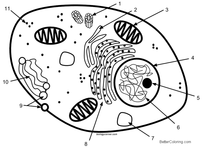
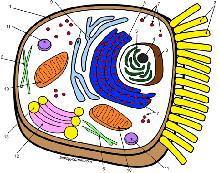
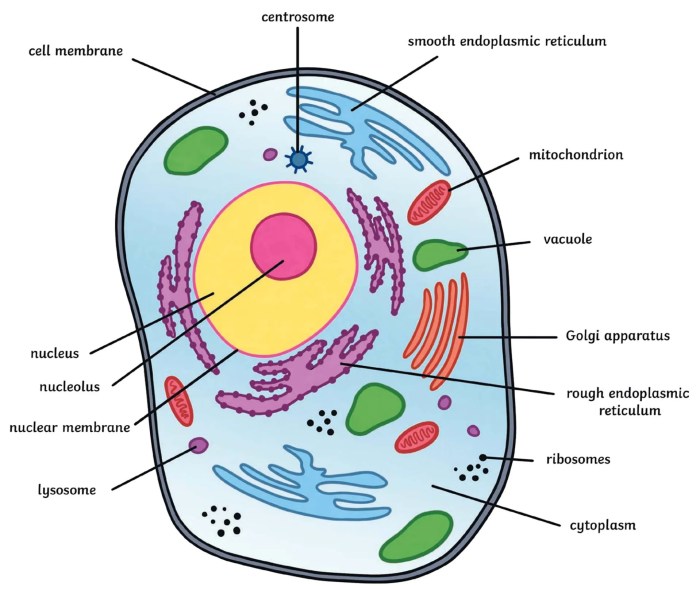
Imagine a perfectly round cell. The outer edge, a light blue line, represents the cell membrane. Inside, a light green jelly-like substance fills most of the space – that’s the cytoplasm. Nestled near the center is a dark purple blob, the nucleus, the cell’s control center. Scattered throughout the cytoplasm are bright red ovals – the mitochondria, buzzing with energy.
Tiny dark brown dots, the ribosomes, are also visible, busily making proteins. A yellow network of interconnected tubes, the endoplasmic reticulum, winds its way through the cytoplasm. Near the nucleus, a stack of flattened sacs, colored orange, is the Golgi apparatus, meticulously packaging proteins. Smaller dark green circles, the lysosomes, are scattered about, diligently breaking down waste. A few light pink sacs, the vacuoles, are visible, storing various materials.
Finally, near the nucleus, small lavender structures, the centrioles, are present, playing a vital role in cell division.
Common Labeling Mistakes to Avoid
Getting organelles mixed up is super easy! Here are some common mistakes to watch out for:Confusing the Golgi apparatus with the endoplasmic reticulum: Remember, the Golgi is a stack of flattened sacs, while the ER is a network of tubes.Misplacing the nucleus: The nucleus is usually centrally located (but not always!).Forgetting the cell membrane: It’s the outermost boundary – don’t leave it out!Incorrectly labeling vacuoles as lysosomes: Vacuoles are for storage, lysosomes for waste breakdown.Failing to distinguish between mitochondria and ribosomes: Mitochondria are larger and oval, while ribosomes are tiny dots.
Understanding animal cell structure can be enhanced through active learning. The process of coloring and labeling an animal cell helps solidify knowledge of organelles and their functions. A helpful resource for this is a visually engaging animal cell coloring page , which can serve as a starting point for your learning. Following this visual exercise, you can then further your understanding by creating your own detailed diagrams, reinforcing the concepts you’ve learned.
Advanced Techniques and Applications

Animal cell coloring and labeling, while seemingly simple, opens doors to a world of advanced techniques and applications crucial in both research and education. Moving beyond basic visualization, these methods allow us to delve deeper into the intricacies of cell biology, revealing dynamic processes and interactions within the cell.Fluorescent Microscopy in Visualizing Animal Cell StructuresFluorescent microscopy is a game-changer in visualizing animal cell structures.
This technique uses fluorescent dyes or proteins that bind to specific cellular components, causing them to emit light at a particular wavelength when excited by a specific light source. The process involves preparing the cells, staining them with fluorescent probes, and then observing them under a fluorescence microscope. The resulting images provide highly detailed and specific information about the location and distribution of various organelles and molecules within the cell.
For example, staining with DAPI (4′,6-diamidino-2-phenylindole) highlights the cell’s nucleus, while other fluorescent probes can target specific proteins, such as microtubules or actin filaments, revealing the cell’s cytoskeleton. The high resolution and specificity of this technique allows researchers to study dynamic cellular processes like cell division, protein trafficking, and signal transduction with unprecedented detail. The results are stunning, vibrant images revealing the intricate workings of the cell in a way that basic staining methods simply can’t match.Applications of Animal Cell Coloring and Labeling in Research and EducationAnimal cell coloring and labeling techniques are essential tools in both research and educational settings.
In research, these methods are used extensively to study a wide range of biological processes. For example, immunofluorescence microscopy, a variation of fluorescent microscopy, uses fluorescently labeled antibodies to detect specific proteins within cells. This technique is crucial for studying protein localization, interactions, and dynamics. Researchers might use this to study the distribution of specific receptors on the cell membrane or track the movement of proteins within the cell during cellular signaling.
In education, these techniques provide a powerful way to visualize and understand complex cellular structures. Students can learn about the different organelles and their functions by directly observing them under a microscope after staining. For instance, observing a cell stained with eosin and hematoxylin allows for clear differentiation of the nucleus and cytoplasm, providing a visual confirmation of their respective functions in protein synthesis and cellular processes.
The hands-on experience of preparing and observing stained cells significantly enhances learning and understanding of cell biology.Creating a Key for Interpreting a Colored and Labeled Animal Cell DiagramA key, or legend, is essential for correctly interpreting a colored and labeled animal cell diagram. The key should clearly list each color or label used in the diagram and specify the corresponding cellular structure or component.
For example, a key might indicate that purple represents the nucleus, blue represents the endoplasmic reticulum, and green represents the Golgi apparatus. A well-designed key should be simple, clear, and easy to understand, using concise descriptions and consistent color coding. It should be placed near the diagram for easy reference. Consider including the magnification level of the image in the key as well, to provide context for the size of the structures shown.
A properly constructed key ensures that anyone viewing the diagram can accurately interpret the information presented.
Practical Exercises and Activities
Yo, Jogja squad! Ready to dive into some hands-on learning about animal cells? This section’s all about practical exercises that’ll make understanding animal cell structures a breeze. We’ll cover a simple coloring and labeling activity, a worksheet design, and how to assess your work. Get your coloring pencils ready!This section details a simple yet effective animal cell coloring and labeling activity, a sample worksheet design, and a method for assessing student understanding.
The activities are designed to be engaging and reinforce learning.
Animal Cell Coloring and Labeling Activity
This activity provides a step-by-step guide for a fun and educational animal cell coloring and labeling exercise. The materials required are easily accessible and the process is straightforward, making it suitable for various learning environments.Materials Needed:
- Worksheet with a pre-drawn diagram of an animal cell (see Worksheet Design section).
- Colored pencils or crayons.
- Pen or pencil for labeling.
- A list of animal cell organelles and their functions (provided by the instructor or sourced from a textbook).
Procedure:
- Carefully examine the provided diagram of the animal cell. Note the different organelles present.
- Using the provided list or your textbook, identify the function of each organelle.
- Choose different colors to represent each organelle. For example, you might use purple for the nucleus, green for the chloroplasts (if included), and yellow for the mitochondria. Consistency is key!
- Color each organelle according to your chosen color scheme. Make sure the coloring is neat and accurate.
- Using a pen or pencil, neatly label each organelle on the diagram. Write the name of the organelle clearly and concisely near the structure you colored.
Worksheet Design
This section describes a sample worksheet design for the animal cell coloring and labeling activity. The design incorporates a clear diagram, sufficient space for labeling, and clear instructions. This ensures the activity is both engaging and effective.The worksheet should feature a large, clear diagram of an animal cell. The diagram should accurately depict the major organelles: nucleus (with nucleolus), cytoplasm, cell membrane, mitochondria, ribosomes, endoplasmic reticulum (rough and smooth), Golgi apparatus, lysosomes, and vacuoles (although animal cell vacuoles are smaller than plant cell vacuoles).
There should be ample space around each organelle for labeling. Clear instructions should be provided at the top of the worksheet, guiding students through the coloring and labeling process. A space for a student’s name and class should also be included. An example: The nucleus could be depicted as a large, centrally located circle, with a smaller circle inside representing the nucleolus.
Mitochondria could be represented as bean-shaped structures scattered throughout the cytoplasm. The endoplasmic reticulum could be shown as a network of interconnected tubes and sacs. The Golgi apparatus could be illustrated as a stack of flattened sacs.
Assessment of Student Understanding
This section Artikels a method for assessing student understanding of animal cell structure based on their completed coloring and labeling worksheets. The assessment focuses on accuracy, neatness, and the demonstration of understanding of organelle function.Assessment will be based on several criteria:
- Accuracy of Coloring: Did the student correctly identify and color each organelle?
- Accuracy of Labeling: Did the student correctly label each organelle?
- Neatness: Was the work presented neatly and clearly?
- Completeness: Did the student complete all parts of the activity?
A rubric can be used to standardize the assessment process. For example, each criterion could be scored on a scale of 1-3 (1=needs improvement, 2=satisfactory, 3=excellent). The total score would then provide an overall assessment of the student’s understanding. Students who demonstrate inaccuracies in coloring or labeling may need additional instruction or review of the material.
